

Table of Contents
Predicting, Preventing, and Managing Complications

As the dental profession continues to define its role in the assessment and management of sleep related breathing disorders, the need to provide education based on research and clinical experience is clearly recognized. Though scientific investigations and technology have provided systems and protocol to diagnose a wide spectrum of sleep disorders, a full understanding of how the profession of dentistry can provide effective therapy continues to evolve.
Snoring, upper airway resistance syndrome (UARS), respiratory effort related arousals (RERA), and obstructive sleep apnea (OSA), have become the focus of dental providers who have developed interest and expertise in sleep disorders. As oral appliance therapy (OAT) designed to advance the mandible has gained a foothold within the therapeutic regimens offered for these conditions as a result of scientific investigations and research, efforts continue to unveil not only best practices, but strategies to limit the potential consequences of using these devices. As a result, careful assessment of the dental structures, jaw muscles and temporomandibular joints from a clinical and imaging perspective is essential before starting OAT.
What You Need to Consider
Before discussing the specifics of the assessment process and how one should go about identifying patients who may be at risk of complications from MADs some general commentary about snoring and obstructive sleep apnea is important. The intent of treatment in patient populations with these problems is to reduce airflow turbulence and facilitate airflow. Snorers without apnea are desirous of a quieter environment for their bed partners. They may also want to reduce the severity of their snoring to spare their airway from the potential structural and neural consequences that have been documented in the literature as a result of long-term airflow turbulence. Beyond these concerns, the patient with OSA is looking for treatment that will reduce or eliminate problematic AHI scores, and prevent persistent oxygen desaturations during sleep. With the knowledge that MADs can put the teeth, jaw muscles and temporomandibular joints at risk, the goal is to maximize benefit and minimize complications over what may be an extended period of time. If, however, despite the best of efforts and practices, dental, occlusal, or jaw problem arise as a result of wearing a MAD, particularly in the patient with OSA, concern must be balanced with recognition of the benefits achieved by keeping the airway patent and preventing oxygen desaturations. In this light, TMD symptoms, open contacts between teeth, or a bite change, may be of little consequence to the patient who has bigger medical concerns. We as dentists must get comfortable with these outcomes.
A Broad Minded Approach
As the complications from OAT are likely the result of forward jaw positioning and it’s influence on the teeth, muscles and TM joint anatomy (disc, retrodiscal tissue) the clinician must always be looking to utilize a broad minded approach to treatment. Continued collaboration with our medical colleagues is therefore essential to employing a combination of treatment strategies to facilitate airflow during sleep. If weight loss is a possibility it should be pursued. If a patient has poor nasal airflow, collaborative ENT consultations and interventions may be in the patient’s best interest, if simple strategies like nasal strips or lavage fall short. Encouraging side sleeping with props should always be a consideration, but certainly used to help the patient avoid the dreaded supine position during REM. Most importantly CPAP therapy and OAT should not be considered as separate unrelated therapies. Marrying these two strategies should be a routine consideration. It should not be uncommon for some of your patients to use CPAP from 10 pm to 3 AM and then if awakened for one reason or another, to put their oral appliance in for the rest of the night if resuming CPAP is objectionable. Others use CPAT and OAT together nightly to help reduce the force of airflow from their CPAP machine. Still others split the week between CPAP and OAT. Lastly, if a MAD is working well, you may want to dial back the appliance position and retest with pulse oximetry or a Home Sleep Test (HST). If a less protrusive position works, the appliance should be reset, reducing the likelihood of complications arising. With these concepts in mind, your ability to help patients will be greatly facilitated.
The Assessment Process – Taking a thorough history
Identifying the patient who may struggle to wear an oral appliance or develop complications down the line is a function of taking a good history. Typical questions that should be asked include;
Did you ever use retainers after orthodontics and were you comfortable wearing them?
Are your teeth presently shifting, particularly across the upper or lower front regions.
Do you have an easily excited gag reflex?
Do you easily develop canker sores, (as this may influence appliance design avoiding metal hardware.)
Do you sleep on your back, side or stomach?
Do you or can you breath through your nose when you sleep or is your mouth open?
Do you clench or grind your teeth at night? As a result of these activities do you have any morning symptoms of tooth soreness, jaw muscle pain, restricted jaw motion, TMJ noises, jaw locking, joint soreness. If so do these symptoms linger or pass quickly?
Have you ever been treated for a TMJ problem? What was the outcome and were your problems focused more in the muscles or TMJs.
Do you currently wear a night guard? Does it help?
Do your TMJs click? Is so, are the noises longstanding? Does your jaw lock or get jammed at any time? Are the noises accompanied by pain?
Are your teeth very sensitive?
Answers to these questions will help determine if OAT is a viable treatment option or whether it may be contraindicated. Information that is communicated up front that pertains to the patient’s ability to wear, be comfortable, and have success with an appliance is viewed as knowledge. Information provided after concerns develop is often viewed as an excuse and is not well received by patients.
The Assessment Process – The Clinical Exam and Imaging
 The place to start is always the teeth, periodontal environment and intraoral tissues. A screening panoramic x-ray should be viewed as the standard of care as it provides a quick glimpse into possible tooth related red flags or contraindications to OAT. Periodontal deficits, and compromised restorations must be identified at the outset, as these problems will require careful clinical scrutiny and may prompt management before the oral appliance is fabricated. The panoramic x-ray will also provide an opportunity to appreciate condylar size and shape that can be correlated with examination findings. Significant alterations in the bony anatomy of the TMJs identified on the panorex may prompt a cone beam scan or MRI if there is clinical evidence of functional limitations. The use of 3D CBCT imaging can independently be utilized to identify airway anatomy that may influence the ability of MADs to assist airflow in patients who have a confirmed obstructive sleep apnea (OSA) based on past PSG and of HST. The use therefore of CBCT technology should be viewed as part of the screening process and not a way to diagnose sleep related breathing disorders.
Intraoral Exam
A thorough intraoral exam will help identify factors that will assist in the process of choosing an oral appliance strategy. For instance, the presence of short teeth that will likely compromise retention of the appliance rims should prompt the use of an interlocking appliance design which limits dislodgement of an appliance when the mouth drops open. Worn teeth with wear facets may indicate a long term sleep bruxism pattern that will prompt the use of specific appliance designs that will help support the TMJs. Anatomy such as narrow dental arches, and a large tongue with scalloping may prompt the selection of an appliance design that allows for small and non-bulky upper and lower rims or a single arch appliance. Encroaching on tongue space often can lead to the onset of jaw pain or intolerance to using the appliance. On the other hand, oversized tonsils (grade3/4) that may predispose to more aggressive forward jaw positioning to open the airway should prompt collaborative medical consultations. Mouth breathers should have their nasal airway assessed to determine if airflow could be improved. If open mouth breathing is the reality of the situation, appliance designs that use elastics to limit mouth opening must be avoided. Evaluating and thinking in this manner can often lead to more effective OAT with less forward mandibular positioning required.
Jaw Function, Comfort and Occlusion
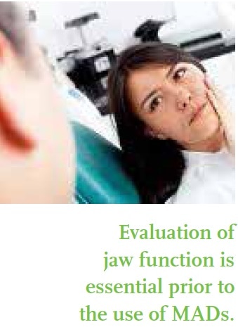
Evaluation of jaw function is essential prior to the use of MADs. Normal jaw motion occurs with fluidity in a smooth, silent and straight path. Jaw range of motion for men is approximately 40-50 mm and for women in the 35-43 mm range. Lateral motions are typically 9-13mm. All motions should occur without pain and the absence of TM joint noises. When measuring protrusion, this is a good time to use a gauge, (e.g. George Gauge or Pro Gauge) which will allow measurement of the full extent of retrusion and protrusion. This information is essential in determining whether the patient has the structural capacity to bring the jaw forward enough to justify OAT. At times in patients with jaw relationships that establish edge-to-edge incisal relationships, there is minimal forward positioning potential of the lower jaw. In these cases using the oral appliance to prevent the tongue from falling back as opposed to bringing the jaw forward may be the best that can be achieved. Once the full extent of retrusion-protrusion is determined, the initial bite position can be set (usually somewhere between 50-60 % of the maximum forward potential), and captured with a bite registration.
Palpation of the jaw muscles will often reveal the presence of hypertrophy in the masseter and or temporalis region with no associated soreness or pain. As over built but non- symptomatic muscles are likely the result of overuse day behaviors (including clenching) and sleep bruxism, these factors must be considered when choosing a MAD. Unfortunately, there are clenchers and grinders in this world who never develop jaw symptoms until a MAD is placed to address an airway problem. This potential complication can best be avoided by careful appliance design that will be discussed under the section on complications. If muscle or joint soreness is discovered, but without daily symptoms, the same overuse factors may be responsible, and similar appliance designs considered. If grinding tendencies (lateral bruxism) are identified while sleeping, the appliance design chosen must not only protect the TMJs but be capable of withstanding nightly forces which can be of significant magnitude.
Joint noises if present without pain or marked pathway alterations may represent a common deviation from normal (Estimated that 30% of the US population experiences joint noises), and may not impact your choice of appliance design. Joint noises that are accompanied by pathway deviations and intermittent jamming or locking events should raise red flags as well as any patient who reports a history of a TMD problem, especially if there was joint pain or instability. A regimen of formal jaw therapy may be required to prepare these patients for OAT inclusive of exercises, formal physical therapy, cessation of daytime overuse behaviors that may be overworking the jaws, and/or a bruxism guard for a period of time. Despite these efforts some of these patients remain poor candidates for OAT. At best it may be possible to use OAT in these patients simply to prevent jaw retrusion.
 Assessing the dental occlusion prior to OAT is essential. This is the time to clinically evaluate the patient’s habitual centric occlusion position and chart contact zones that if altered will prompt an awareness of a changing bite. With today’s technology, photographs along with digital representations of the occlusion should be included in the record for future reference. Study models and or elastomeric impressions that remain stable can also be used for this purpose.There does not appear to be any one specific occlusal relationship that will predispose to a bite change in the morning, nor are there any readily identifiable occlusal factors that will consistently predispose to either transient or long term bite changes. Patients, however, who start with habitual bites that have variable contact zones seem to develop changes more readily, as evidenced by premature anterior contacts and a reduction of the grip of posterior contacts on articulating paper. Patients with lingually inclined maxillary incisors and deep bites may also develop this change more readily but not predictably. The presence of heavy and premature anterior contacts and a reduction of gripping contacts posteriorly or a posterior open bite is the typical scenario with most bite changes that can occur as a result of MADs.
Complications of MADs
 As previously mentioned, mandibular advancement devices (MAD) are presently used with the intent of moving the lower jaw and tongue base forward and or preventing the tongue from moving back into the oropharynx. This specific action reduces the collapsibility of the airway, minimizing or preventing snoring and/or airflow compromise leading to OSA. Forward jaw posturing, maintained over several hours repeated daily, however, is not normal, and can lead to symptoms in the teeth, jaw muscles or TMJs. The extent to which the oral appliance must move the mandible forward in order to achieve the designated goals is a point of important consideration and will likely influence the development of complications over time.
Complications that occur as a result of using OAT include; tooth sensitivity, tooth movement leading to open interproximal contacts and anterior interferences, muscle stiffness and soreness, joint pain, the onset or escalation of joint noise, and changes in the way the teeth articulate. For most patients these problems are minor, transient in nature and easily managed. There are, however, risk factors that can increase the potential for complications and or make them more difficult to manage. As a result, identifying a history of a TMJ problem, ongoing nocturnal clenching or lateral bruxism, and patterns of nasal/mouth breathing while sleeping are invaluable to the clinician to assist in choosing a specific appliance design. Though all oral appliances to address OSA are intended to bring the jaw forward or prevent the tongue from falling back, there are a number of designs. By identifying risk factors at the outset, the clinician is in a position to choose a design that will afford the greatest protection to the teeth and jaw structures. The careful clinician becomes familiar with the various designs available, so that patient-appliance matching can be as predictable as possible.
The Teeth
 Tooth sensitivity is the most likely symptom due to stress that is placed on the periodontal ligaments. The upper and lower anterior teeth are the most common site of discomfort/pain especially if an anterior stop appliance is used. Reducing the extent of facial surface coverage is at times all that is needed to address these complaints. At other times relief of the internal aspect of the appliance is helpful particularly if an all-acrylic appliance is used. Laminate appliance designs with a soft inner lining often limits tooth sensitivity but may be more contributory to open contacts developing in the premolar and molar region. Frequent floss checks by patients are essential to identify this concern early on. At times tooth shift anteriorly can occur, but usually in periodontally compromised people. A component of the examination assesses potential tooth anchorage for anteriorly posturing the mandible; if suspect, careful appliance choice to distribute force and minimize stress put on individual teeth is even more important.
Muscle Problems
Muscle symptoms of soreness and stiffness can occur in the masseter region particularly when OAT is first initiated. This may be the result of muscle bracing occurring in the awkward forward jaw position or the result of contact of the occlusal rims creating masseter contraction even without defined bruxism. If the appliance chosen is too big and obliterates freeway space (evidenced by loss of lip seal), the patient is almost forced to clench with possible pain symptoms emerging. Changing the appliance design would therefore be required. Muscle symptoms, however, are more profound in the patient who clenches or grinds their teeth. For these patients using an appliance that provides two broad posterior contacts and anterior contact without obstructing the anterior opening is desirable. The Herbst, Suad, Somnodent, Respire and EMA can all be used in this situation with predictability. The use of an anterior point contact design may over time load the anterior compartment of the TMJ so this would not be an ideal option, unless posterior contact pads are added. In general, a hot shower and repeated circular massage of the masseters for six seconds followed by six unassisted jaw opening movements is usually sufficient to address sore masseters in the morning. One specific isometric exercise can be used as well: Have the patient lock the tip of their tongue against the palate and have them open their mouth while placing the thumb under the chin for resistance. Hold this resistance for 6 seconds and repeat six times. This will relax the temporalis and masseter muscles. If specific appliance designs plus morning exercises falls short, then injection of between 50-100 units of Botox can be considered. Though this may seem excessive, in a patient with OSA that cannot tolerate CPAP or benefit or chose surgery, making an oral appliance work is the ultimate goal.
For the lateral bruxer, posterior contact supports are desirable, but avoid dorsal fin designs, along with Herbst and Suad devices as the upright wings and metal arms respectively can break. The aggressive lateral bruxer typically does well with an EMA appliance as long as posterior contacts are built into the design. The elastic straps will require frequent change as they will stretch and allow the jaw to slip back. The TAP 3 Elite, which allows lateral shift anteriorly, is a reasonable option as long as posterior occlusal pads are built into the design.
Joint Problems
TM joint problems are often difficult to manage and may require a period of time during which the appliance is discontinued. As with all orthopedic joint systems, ligament sprains can occur with and without inflammation. Sprains can occur simply as a result of forward jaw positioning with the MAD, but can be a consequence of clinician error as well. If the centric occlusion midline position is violated as the mandible is advanced, joint sprains can occur. Care must be taken during bite registration to avoid this complication. Once initiated, joint sprains and inflammation may be stubborn to resolve. If a three or four day regimen of NSAIDS doesn’t solve the problem along with a thoughtful diet and some ice massage, there may be good reason to reduce the forward positioning. As most of the appliances used to address a mild to moderate OSA problems are initially set at a position that represents 50-60% of maximum protrusion, there is ample room to back track and allow for healing to occur. If a period of time without the appliance is required this is an option that may have to be considered particularly if a patient has a CPAP machine that they can use. A tongue-repositioning device (Aveo tongue stabilizing device or MPowRX) can also be tried for a short period of time until the joint has healed. When symptoms have passed and appliance therapy is reinitiated, an every other night approach with less than optimal positioning should be pursued for a period of time. There have been times when a short regimen of physical therapy has helped a sprained joint to recover allowing the resumption of effective OAT. The potential for joint problems to occur and linger increases in the lateral bruxer over time.
Occlusal (bite) changes that are mild to profound appear to occur in approximately 20% of patients that are followed beyond one year. For the dentist newly involved in OAT this is a dreaded complication. For the experienced dentist and patient with OSA, this consequence, if explained before treatment was started, is a mere trade off with the benefits being realized in reduced blood pressure, weight loss, gastrointestinal health, refreshing sleep and improved daily energy levels to name just a few positive outcomes. There are a number of strategies and techniques that can be employed to address these anticipated bite changes. The most useful requires obtaining a durable centric occlusion bite registration prior to OAT. This can be done with strong elastomeric materials or baseplate wafers. The AM Aligner from Airway Management has worked exceptionally well, particularly if you cut back the length allowing the second molars to contact naturally and the rest of the bite to captured in the baseplate wafer. In the morning, the patient puts the Aligner in their mouth and while in a hot shower, bites into the bite registration with moderate effort over a three to five minute period of time. This coupled with some gentle circular massage of the masseter and temporalis muscles usually resets the centric occlusion position. If this falls short, gentle gum chewing while getting dressed helps fully recapture the bite. If needed, employing an isometric exercise can be helpful. Have the patient open their mouth halfway and then close against resistance provided by the index and middle finger for 6 seconds, and then repeat 6 times. This isometric exercise should assist relaxation of the lateral pterygoid muscles that may be guarding after hours of forward jaw posturing. Despite these efforts it may take until mid morning for the bite to settle back to normal. If this is the scenario, it is likely that the morning bite change may relate to shape changes of the articular disc or retrodiscal tissues as a result of the condyles being moved forward for several hours. This often is a harbinger of more profound bite changes occurring over time and may be inevitable.
Conclusion
As OAT provided by dentists to address snoring and obstructive sleep apnea gains more public visibility and acceptance by our medical colleagues, standards of care and best practices will need to evolve based on research and clinical experiences. Though MADs have proven to be effective and embraced by patients looking for alternatives to surgery or CPAP therapy, much work needs to be done. Future efforts should focus not only on how to more predictably determine the start position for MADs but to establish systems and strategies that allow patients to use these devices over extended periods of time with minimal complication.