The use of the Cone-beam Computed Tomography (CBCT) of the maxillofacial region has quickly created a revolution in all fields of dentistry. It has expanded the role of imaging from diagnosis to image guidance for many procedures and has eliminated some of the inherent limitations of 2D images such as magnification, distortion, superimposition and misrepresentations.
Image Selection
The American Academy of Oral and Maxillofacial Radiology (AAOMR) has established the groundwork and guidelines for image selection in diagnosis, treatment planning and follow up of a patient with conditions affecting the TMJ. Conventional radiographic TMJ projections like transpharyngeal, transcranial, panoramic radiograph, and tomographic sections of TMJ may be adequate in a number of clinical situations. However, there are bony alterations that occur in these disorders like erosions, osteophytes, and pneumatisation of the articular eminence that are difficult to detect in conventional radiographs due to overlapping of the anatomic structures. Because of this, the use of advanced imaging modalities like Magnetic Resonance Imaging (MRI), arthrography, conventional Computed Tomography (CT) and CBCT are warranted.
Let’s take a look at a few of the advanced imaging modalities and their use in diagnosis and treatment:
MRI: Imaging of the TMJ should be performed on a case by case basis depending upon clinical signs and symptoms. An MRI is the diagnostic study of choice for evaluation of disk position and internal derangement of the joint because it provides high resolution and great tissue contrast.
Arthrography: Today, arthrography is rarely used because an MRI can be used to evaluate the TMJ without being invasive, exposing the patient to a possibility of allergic reaction from the contrast, the possibility of infection, or using radiation.
CT Scan: The use of a CT scan for evaluation of TMJ is indicated if bony involvement is suspected. This choice should be judiciously considered because of radiation exposure.
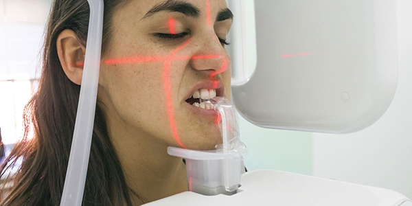
CBCT Imaging: By utilizing CBCT imaging, clinicians receive sub-millimeter spatial resolution images of high diagnostic quality with relatively short scanning times (10–70 seconds) and a reported radiation dose equivalent to that needed for 4 to 15 periapical radiographs. CBCT imaging is ideal for the evaluation of fractures, degenerative changes, erosions, infection, airway volume, sinus, nasal passages as well as congenital anomalies.
Evaluation of the airway has become an important diagnostic test in several subspecialties of dentistry. Multiple orthodontic researchers have developed techniques to use full-head x-rays to determine airway obstruction, and the potential impact of high-resistance airways leading to abnormally developed increases in the vertical facial dimensions in young patients. CBCT imaging has opened up the opportunity to evaluate the cross-sectional area of the airway as well as the volumetric 3-dimensional (3D) depiction of the entire airway using a lower-radiation method than a medical CT. The CBCT system provides a low-radiation, rapid- scan capability to assess patients’ airway using a highly correlative linear, cross-sectional area, and volumetric measurements that include assessing the morphometry of the airway.
Guidelines
Clinicians must abide by the ALARA (As Low As Reasonably Achievable) principle when ordering an imaging modality for a patient. Exposing the patient to the radiation must provide an image whose diagnostic value is greater than the detriment the radiation exposure may cause. Not every patient requires a CBCT because it does expose the patient to radiation and results in increased cost. The ADA Council on Scientific Affairs also suggests that CBCT use should be based on professional judgment and clinicians must optimize technical factors such as using the smallest field of view (FOV) necessary for diagnostic purposes and using appropriate personal protective shielding.
The ADA Council on Scientific Affairs also suggests that CBCT use should be based on professional judgment
Imaging for TMD and sleep has risen to a new level for TMD treatment, airway analysis and dental implants. Many of the scans are considered medically necessary in my practice which is limited to TMD treatment and Dental Sleep Medicine and are billed through medical insurance.
For more related education, dentists may visit my office in Atlanta, GA as part of our “Shadow a TMD and OSA Dentist Program” through Nierman Practice Management CE or attend a seminar related to TMD and dental sleep medicine. For more information, contact Jon Nierman at 800-879-6468 or visit www.NiermanPM.com.